
PERIPHERAL ANGIOGRAPHY
PERIPHERAL ANGIOGRAPHY
Peripheral angiography is a minimally invasive imaging procedure used to examine the blood vessels in your arms and legs. It’s a two-part process that allows doctors to diagnose and sometimes treat blockages in these peripheral arteries.
Diagnosis:
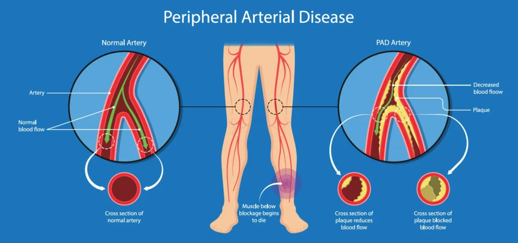
- Peripheral artery disease (PAD): the most common reason for an angiogram. PAD causes narrowing of the arteries due to plaque buildup, reducing blood flow to the legs and feet. Symptoms include leg pain, cramping, sores that won’t heal, and coldness.
- Aneurysms: weak bulges in the artery wall that can rupture and cause life-threatening bleeding.
- Blood clots: can block blood flow and cause tissue death (gangrene).
Procedure:
- Catheter Insertion: A thin, flexible tube (catheter) is inserted into an artery in your groin, wrist, or arm.
- Contrast Injection: A contrast dye is injected through the catheter into the target arteries. This dye shows up clearly on X-ray images, highlighting the blood flow and any blockages.
- X-ray Imaging: X-ray images are taken as the dye travels through your arteries. These images provide a detailed view of the blood vessels and any abnormalities.
IR Treatment During Angiography:
Interventional radiology (IR) uses imaging techniques like X-rays during angiography to perform certain treatments on the blood vessels. Here are some IR treatments that can be done concurrently with an angiogram:
- Angioplasty: A tiny balloon catheter is inserted and inflated at the blockage to open the narrowed artery.
- Stenting: A small mesh tube (stent) is placed in the artery to keep it open after angioplasty.
- Thrombolytic therapy: Clot-busting medication is delivered directly to the clot through the catheter to dissolve it and restore blood flow.
Benefits of Peripheral Angiography with IR Treatment:
- Minimally invasive compared to open surgery.
- Quicker recovery time.
- Can diagnose and treat blockages in one procedure.
- Improves blood flow and relieves symptoms of PAD.